Microarray Reader – Microarray Imager – Microarray Scanner
Microarray scanner, microarray reader and microarray imager are terms often used interchangeably to refer to the same type of instrument, but they can also carry slightly different connotations depending on the context. Generally, both microarray reader and microarray imager terms refer to instruments designed to capture and analyze the signals emitted from microarrays. However, there might be nuances in their usage:
Microarray Scanner: This term is commonly used to describe instruments that are specifically designed to scan microarrays and capture the fluorescent signals from the labeled samples on the microarray surface. Microarray scanners usually consist of a scanning mechanism that moves a laser or other light source across the microarray slide, while detectors capture the emitted fluorescence. The term “scanner” reflects the process of moving across the microarray slide to scan the entire surface systematically. A microarray scan is intrinsically slower than an image, but may yield higher resolution.
Microarray Imager: This term, or microarray reader, is often used to describe instruments that capture and analyze images of the microarrays. The term “imager” emphasizes the capturing of images in a single event, rather than just scanning, suggesting a more comprehensive process that involves both capturing and processing image data.
axiREADER
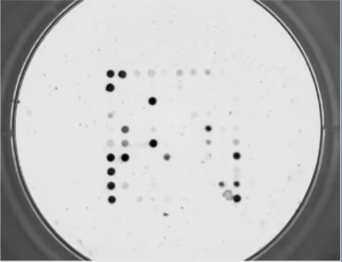
The axiREADER is a microarray imager (microarray reader). It is equipped with specific optics that can detect and quantify the signals generated on a microarray slide or microarray in a well. These signals are typically produced by labeled DNA, RNA, or protein samples that have been hybridized to specific probes on the microarray surface. The imager captures the emitted fluorescence or color intensity and converts it into digital data that can be processed and analyzed using software.
The imager’s performance, such as sensitivity, resolution, and dynamic range, is crucial for obtaining accurate and reliable data from microarray experiments. The axiREADERs have a resolution of 6 um/pixel. They are thus suitable for the analysis of microarrays where spot sizes are 80 um or greater.
Colorimetric vs Fluorometric
Performing multiplex tests and microarrays experiments require a detection step, usually with Microarray imagers. Two of the most commonly used detection modalities for microarrays are colorimetric and fluorometric. The axiREADER microarray reader is available as colorimetric or fluorescence versions. The compact and portable instruments can image microarrays from microtiter plate wells or from slides.
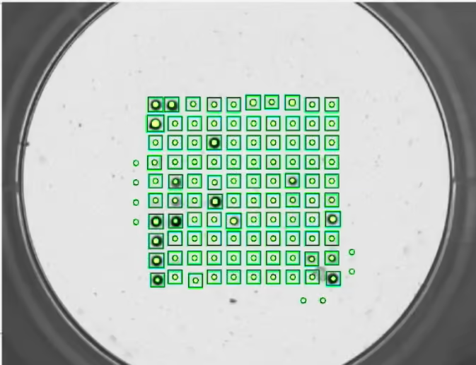
The software uses artificial intelligence for the best in automated spot finding, and quantitative data analysis. Many powerful features are built-in to allow complex data analyses. It can use data from plate level wells, in addition to intra-well arrays values, to perform calculations. For example, your test may include standard calibration curves with the target analyte varying concentrations in each well. This is standard good assay design practice and the axiREADERS can fully accommodate the data set.
axiREADER IMAGERS:
The axiREADERS microarray readers are available as colorimetric or fluorescence versions. The compact and portable instruments can image microarrays from microtiter plate wells or from slides. You can get information on imagers vs scanners here. The instruments can work with a 96-well plate, a 12 x 8 strip well plate, as well as standard 25 x 75 mm slides. Other devices can be read using custom adapters. The microarray imager has an automatic mechanism for tray loading and unloading.
FEATURES:
- Open OEM Platform: YOUR LOGO, NO ROYALTIES
- Robust modern design desktop microarray imager
- High-throughput analysis
- Flexible formats: plates and slides are STD, other format via a custom adapter
- Designed and priced for multiple units deployed for diagnostics and other multiplex analyses
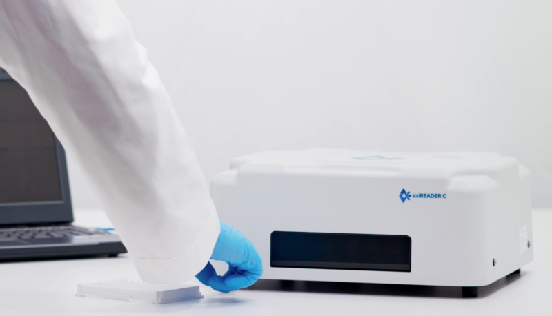
COLORIMETRIC Microarray reader – axiREADER C
The colorimetric imager designed for imaging arrays of biomolecules (after visible staining such as with TMB substrates for example).
The device takes images from above with a highly sensitive CMOS camera capable of taking images in a short exposure time. Illumination from above or below means that surfaces can be either opaque, as for nitrocellulose or transparent, as for glass slides or plastic microtiter plates.
FLUORESCENCE Microarray reader – axiREADER F
The fluorescence imager designed for red/green imaging arrays of biomolecules using 2 or more channels. CY3 and CY5 channels are included in the base system. Readers with 3 or 4 channels are also available.
The device takes images from below with a highly sensitive CMOS camera capable of taking images in a short exposure time. Excitation is also from below which means that surfaces where the array is spotted must be transparent. The use of black ‘chimneys’ for microtiter plates is recommended but not necessary. Alternatively, as for imaging iRiS chips, the substrate can be loaded upside down.
| Features | Specification |
|---|---|
| Light source | White LED |
| Illumination | Brightfield and Darkfield (bottom and top illumination) |
| Lifetime of light source | > 10,000 hours |
| Certified OD measurement range (for brightfield) | OD 0.1 to 2.0 |
| Certified Diffused reflectance meas. range (for darkfield) | 2 to 99 |
| Features | Specification |
|---|---|
| Light source | Laser (optional specific lasers available |
| Illumination | from below (bottom illumination) requires transparent material. |
| Lifetime of light source | > 10,000 hours |
| Fluorescence channels | CT3 and CY5 (basic system) - 3ch or 4ch optional |
| Exposure | Up to 30 sec |
| Features | Specifications in common |
|---|---|
| Vessels | 96 well plate / 12×8 well strip / 4 microscope slides, and more with custom inserts |
| Camera | CMOS, 3 MP |
| Resolution | 6 um per pixel |
| Magnification | > 10X |
| Focus | Manual or Automatic (controllable) |
| Arbitrary measurement range | 0 to 65,535 |
| Imaging speed | 2 to 3 min per 96-well plate (depending on focus mode) |
| Software | Instrument control, image processing, data analysis with algorithms. All included. |
| Import sample file formats | GAL or Excel |
| Export data formats | Excel, csv, PDF, Word |
| Export image formats | PNG, TIFF, BMP, JPG (16-bit) |
| PC requirements | Intel I7 or equivalent, 8GB RAM , 256 GB SSD, Win 10 or 11 (64-bit), Nvidia GeForce 10/16/20 (only) |
| PC interface | 1x USB 2.0 and 1x USB 3.0 SuperSpeed |
| Unit dimensions | 330 x 345 x 150 mm (6 kg), 13 x 13.6 x 6.7 in (15 lbs) |
| Power | 20V, 2.5A, 50 W - can be battery operated |
| Power supply | Input 100–240 VAC 50/60 Hz, Output 20-24 VDC, 2.5A |
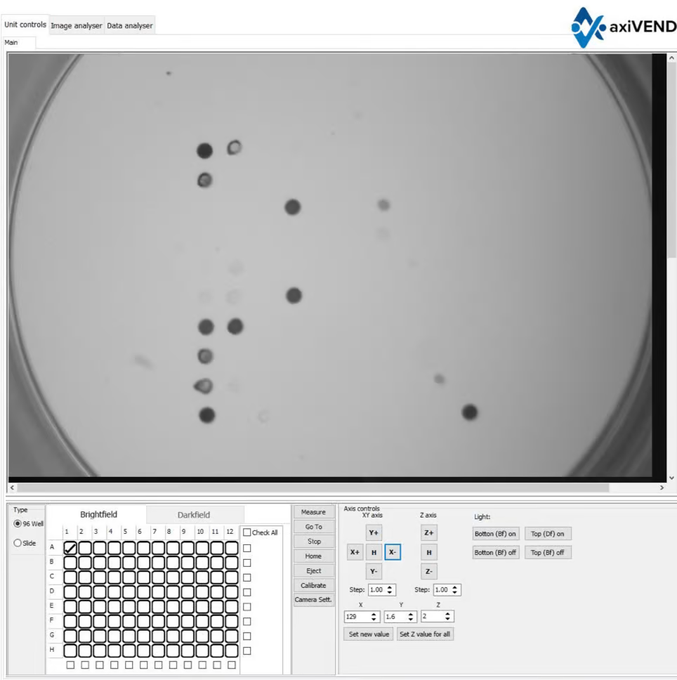
UNIT CONTROLS FEATURES:
- Brightfield and darkfield illumination (Bottom and Top)
- XYZ axis control, Z for focusing
- Specify measurement locations
- Calibrate measurement locations for 96 well plate or 4 microscope glass slides
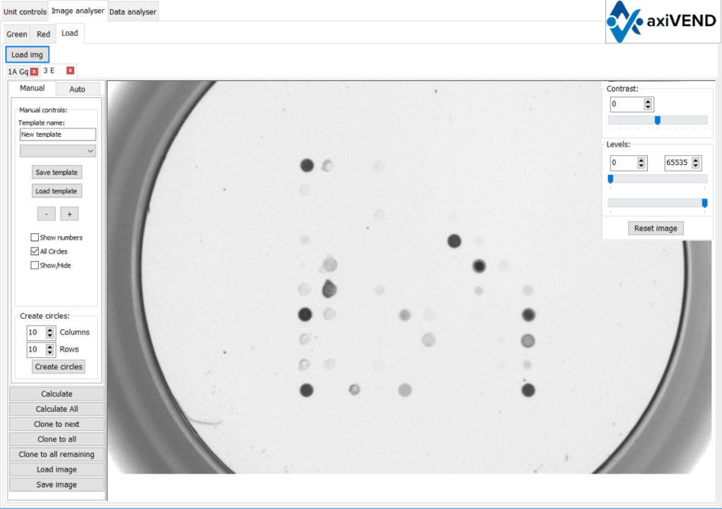
IMAGE ANALYSIS OPTIONS:
- Manual control of the array’s grid positioning
- Individual control of every spot
- Microarray image processing
- Creation of template arrays
- Saving/Loading array’s
- Automated spot finding
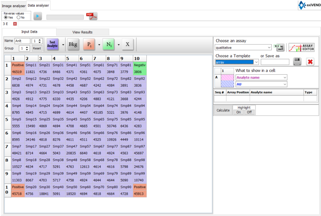
DATA ANALYSIS:
- Select qualitative / quantitative assay for interpretation
- Set the template of your analytes and controls
- Set names for analytes
- Set replicates of analytes /controls
- Save, load and export results
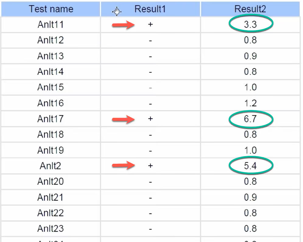
- Creates visual reports (Excel, PDF, CSV)
- Custom reports available
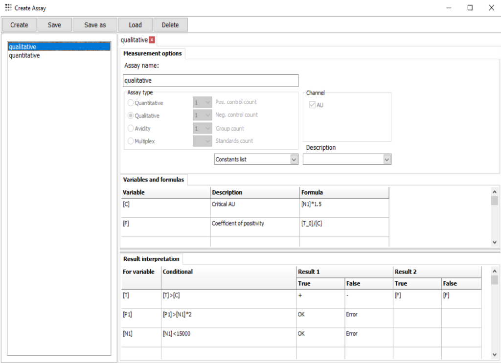
ASSAY EDITOR:
- Qualitative assay with up to 11 control groups (pos/neg)
- Quantitative assay with up to 20 standards
- ‘BestFit’ function for selecting the best calibration curve
- Set individual variables and formulas for analytes and controls
- Create analyte-specific algorithms for data interpretation
- Set the quality control interpretation for your assays
