

Microarray imaging and analysis:
Microarrays are powerful tools used in genomics and proteomics research to analyze the expression levels of multiple genes or proteins simultaneously. They allow researchers to study expression patterns, identify biomarkers, and gain insights into various biological processes. The imaging and analysis of microarrays involve several steps.
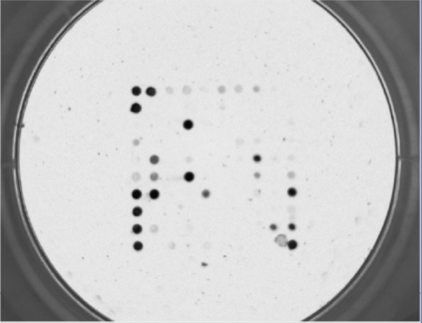
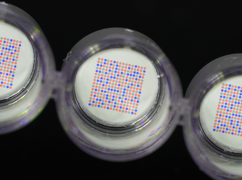
Microarray imaging: The microarray is either scanned or imaged using a specialized reader that excites the fluorophores or detection molecules and captures the emitted signals, or captures the color intensity of the various spots. The reader detects the fluorescence intensity at each spot on the microarray, generating an image of the microarray.
Microarray analysis: The acquired microarray image is then analyzed using bioinformatics tools and software. In the case of our axiREADERs, the builtin software is used for analysis. The analysis involves several steps, including spot finding, signal quantification, and normalization. Spot finding algorithms identify the positions of the spots on the microarray, while signal quantification determines the fluorescence intensity at each spot. Normalization methods are applied to account for technical variations and normalize the data across multiple samples.
Microarray data interpretation: Once the microarray data is analyzed, it can be further processed and interpreted. Researchers may perform statistical analysis, clustering, or other bioinformatics techniques to identify differentially expressed genes, patterns of gene expression, or other relevant information.
This process is fully implemented in our line of axiREADERs.
